
Newborn Nursery at Lucile Packard Children's Hospital
Excellent Care from the Moment of Birth
Stanford Medicine News about Pediatric Care and Research
-
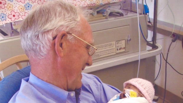
Alistair Philip dies at 86
Alistair Philip, professor emeritus of pediatrics, pioneered a test to reduce antibiotic use in newborns, streamlined nursery care at several hospitals and devoted his life to educating others in his field.
-
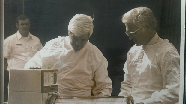
Alvin Hackel dies at 91
The Stanford Medicine professor emeritus of anesthesiology and of pediatrics invented a transport incubator for newborns and helped establish pediatric anesthesiology as a specialty.
-

Less sleep, activity linked to prematurity
Data from wearables show that deviations from normal sleep and activity in pregnancy are connected to a risk for premature delivery, a Stanford Medicine-led study found.
Expectant Parents
For information about maternity services at Lucile Packard Children's Hospital, please visit the Johnson Center for Pregnancy and Newborn Services website.
About this Site
Part of the Johnson Center for Pregnancy and Newborn Services, we specialize in the medical evaluation and care of newborns who are at or near term.
We designed this site to be a resource for our pediatric trainees as well as health professionals worldwide who care for newborns.
The transition from life in the womb to the outside world is an amazing process. We are priviledged to care for babies as they make this transition and adapt to their new surroundings.